Introduction
Thyroidectomy is a common surgical procedure in the world, and is highly prone to complications since it is carried out in a narrow anatomical area where many vital anatomical structures are located [1].
One of the most important and the most feared complications of thyroid and parathyroid surgery is recurrent laryngeal nerve injury. Recurrent laryngeal nerves are located lateral to the ligament of Berry and behind the tubercle of Zuckerland where they enter the larynx bilaterally. Various anatomical alteration in course of recurrent laryngeal nerves has been reported including non-recurrent course. Recurrent laryngeal nerve injury has been reported as 0.6-1% even in experienced hands. Therefore it is important to identify the course and protect recurrent laryngeal nerve [2].
Injury to the recurrent laryngeal nerve leading to vocal cord paralysis is one of the most serious complications seen after thyroid surgery. The average incidence of temporary postoperative vocal fold palsy is around 3.8%.3 When it is bilateral it may cause major respiratory consequences requiring tracheostomy or segmental posterior cordectomy. Surgeons must have a comprehensive understanding of the anatomy of RLN during thyroid operation. There are several anatomical variations of nerve ranging from extra-laryngeal branches, distorted pattern, intertwining between branches of the recurrent laryngeal nerve and the inferior thyroid artery & non-recurrent laryngeal nerve which might increase the risk of iatrogenic damage.
In this case report, we present a rare intraoperative finding of a double recurrent laryngeal nerve identified on the left side during total thyroidectomy for a large bilateral benign multinodular goitre. Recognition of this uncommon anatomical variation and meticulous dissection allowed safe preservation of both nerve components which is important from the perspective of preventing postoperative vocal cord dysfunction.
Case presentation
A 69-year-old male presented with a progressively enlarging anterior neck swelling involving both sides of the neck. He first noticed a small, painless swelling on the right side of the neck approximately 10 years back. The swelling grew in size gradually and approximately four years prior to admission, it had enlarged to the size which started bothering the patient. He sought medical consultation however reportedly no intervention was advised at that stage as he remained otherwise asymptomatic.
One month prior to admission, the neck swelling increased markedly in size and extended towards the left side. This prompted further evaluation at a secondary care center. Magnetic resonance imaging (MRI) of the neck and fine needle aspiration biopsy (FNAB) were performed. Cytology revealed no malignant cells. He was subsequently referred to Dr. Sardjito General Hospital for definitive management. At the time of admission continued to report bilateral neck swelling without pain or discharge. The patient had no known history of diabetes mellitus, hypertension, allergies, chemotherapy and no significant family history of any thyroid disorder was reported. On initial assessment, the patient was clinically stable with a temperature of 36°C and oxygen saturation (SpO₂) of 99% on room air. Pupils were found to be bilaterally equal (3 mm) with intact light reflexes. No features suggestive of exophthalmos were observed. On inspection a lump was clearly visible on both the left and right sides of the neck with the color similar to the surrounding skin and no wound was present. On palpation, lumps were palpable on both sides of the neck and the masses moved with swallowing. Baseline hematologic, renal, and hepatic parameters were within normal limits. Thyroid function was euthyroid (TSH 1.66 μIU/mL; FT4 1.16 ng/dL). Random blood glucose was elevated (177 mg/dL), while serum electrolytes were within normal ranges.
Neck X-Ray revealed a right-sided neck soft tissue mass causing leftward deviation of the trachea with luminal narrowing at the C7–T2 level. Lung fields appeared normal, heart size was normal and no skeletal abnormality was seen in the visualized skeletal structures. There was no evidence of cervical vertebral fracture or listhesis. The C5–C6
intervertebral disc space appeared reduced suggesting degenerative disc narrowing. Approximate radiographic tracheal diameter of widest and narrowest portion was approximately 1.90 cm and 1.74 cm respectively (Figure 1).
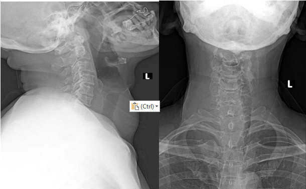
Neck MRI demonstrated a large right thyroid mass with central cystic and subtle degenerative change. These findings raised concern for an underlying neoplastic process. The lesion displaced the trachea and esophagus to the left with associated airway narrowing and appeared to extend into the mediastinum/intrathoracic region. In view of these findings, fine-needle aspiration biopsy (FNAB) was performed. Cytology showed benign features with no malignant cells identified (Bethesda system category II), consistent with a benign follicular (nodular goitre) lesion with chronic inflammatory changes. Overall, the patient was having bilateral suspected non-toxic thyroid nodules (euthyroid multinodular goitre) with compressive effect on the trachea, without clinically evident cervical lymphadenopathy.
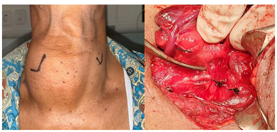
Total thyroidectomy was planned under general anesthesia. During surgery a standard transverse cervical collar incision was made and subplatysmal flaps were raised. The thyroid gland was found to be significantly enlarged bilaterally. Dissection was performed in a capsular plane with careful control of the superior and inferior pedicles. During identification of the recurrent laryngeal nerve in the left tracheoesophageal groove, an extremely rare anatomic variation was noted in the form of two distinct recurrent laryngeal nerve structures (double RLN). They were found in close proximity and ascending toward the laryngeal entry point. Both nerve components were meticulously traced and clearly identified throughout the critical dissection area near the ligament of Berry. Both nerve trunks were separately traced and each entered the larynx independently. Both components were preserved without traction or thermal injury during completion of the thyroidectomy (Figure 2).
Postoperatively patients remained clinically stable with no respiratory distress. There was no hoarseness of voice suggestive of recurrent laryngeal nerve injury. Moreover, there were no features of hypocalcemia during postoperative period. Histopathology confirmed benign multinodular goiter with no malignancy.
Discussion
The recurrent laryngeal nerve (RLN) was first described by Galen in the second century AD who demonstrated its functional importance by showing that transection of the nerve caused loss of voice in experimental animals [4]. In thyroid surgery preservation of the RLN remains one of the most critical steps because injury can result in significant morbidity including hoarseness, aspiration risk and airway compromise. Anatomically, the RLN is the major motor nerve for the intrinsic muscles of the larynx (except the cricothyroid) and also carries sensory and autonomic fibers [5]. It arises from the vagus nerve, looping around the right subclavian artery on the right side and the arch of the aorta on the left, before ascending in the tracheoesophageal groove toward the laryngeal inlet. The course of the RLN may be altered by thyroid pathology and the nerve is particularly vulnerable near its laryngeal entry point.
Safe intraoperative identification of the RLN relies on consistent surgical landmarks and meticulous dissection. Approaching the nerve in a controlled plane and tracing it carefully remains the most reliable preventive strategy [6]. The tubercle of Zuckerkandl has been emphasized as a dependable landmark for localizing the RLN during thyroidectomy with most series reporting the nerve lying medial to the tubercle [7]. In addition the region around Berry's ligament is especially important because the RLN is closely related to this area making careful dissection essential to avoid traction or thermal injury.
The RLN demonstrates considerable anatomical variability. This directly increases the risk of inadvertent injury if the nerve is assumed to be single and unbranched. A large meta-analysis has shown that extra-laryngeal branching is common with an overall pooled prevalence of approximately 60%, meaning that more than one RLN component may be present before laryngeal entry [8] . Another clinically important variant is the non-recurrent laryngeal nerve, a rare embryological anomaly that carries a higher risk of iatrogenic damage if not anticipated and systematically searched for. True duplication of the RLN, however, is extremely rare and mainly described in isolated case reports.
Manoğlu et al reported a left-sided double RLN encountered during thyroid surgery. The authors emphasized that both nerve components must be clearly identified and preserved throughout dissection to prevent postoperative vocal cord dysfunction [9]. Similarly, Mishra and Dhall described a double trunk RLN identified intraoperatively underscoring that such rare configurations can be missed unless the surgeon traces the RLN meticulously up to its laryngeal entry particularly in the region of Berry's ligament where the nerve is most vulnerable [10].
Conclusion
Complete visualization of the recurrent laryngeal nerve (RLN) is important during thyroidectomy to minimize the risk of nerve-related complications. Surgeons must maintain a high level of awareness for possibility of neural variations including the rare occurrence of a double RLN. This is necessary to avoid inadvertent transection or injury to the nerve particularly near the ligament of Berry where the nerve is most vulnerable. Careful capsular dissection, systematic tracing of the nerve up to its laryngeal entry point and preservation of all identified nerve branches remain the most effective measures to ensure patient safety and prevent postoperative vocal cord dysfunction.
Consent and ethics
Written informed consent was obtained from the patient for publication.
References
- Stefanou CK, Papathanakos G, Stefanou SK, Tepelenis K, Kitsouli A, Barbouti A, Tsoumanis P, Kanavaros P, Kitsoulis P. Surgical tips and techniques to avoid complications of thyroid surgery. Innov Surg Sci. 2022 Oct 11;7(3-4):115-123. doi: 10.1515/iss-2021-0038.PMID: 36561510; PMCID: PMC9742273.
- Yadav K. Double Right Recurrent Laryngeal Nerve:Intraoperative Identification. J Oncology. 1st ed. 2022;2(2).
- Ray CS, Sahoo KA. Duplication of recurrent laryngeal nerve in thyroid surgery: a rare anatomical variation. J Oral Med, Oral Surg, Oral Pathol, Oral Radiol.2020;6(2):85–8.
- Kaplan EL, Salti GI, Roncella M, Fulton N, Kadowaki M. History of the recurrent laryngeal nerve: from Galen to Lahey. World J Surg. 2009;33(3):386–393.
- Wojtczak B, Kaliszewski K, Sutkowski K, Bolanowski M, Barczyński M. A functional assessment of anatomical variants of the recurrent laryngeal nerve during thyroidectomies using neuromonitoring. Endocrine. 2018 Jan;59(1):82-89. doi: 10.1007/s12020-017-1466
- Kastan OZ, Ozturk S, Calguner E, Agırdır BV, Sindel M. Relationship of Recurrent Laryngeal Nerve with Inferior Horn of Thyroid Cartilage, Berry's Ligament and Zuckerkandl's Tubercle. Indian J Otolaryngol Head Neck Surg. 2022 Oct;74(Suppl 2):2065-2070. doi: 10.1007/s12070-020-02018-1. Epub 2020 Aug 2.
- Gurluler E. The use of Zuckerkandl's tubercle as an anatomical landmark in identifying recurrent laryngeal nerve and superior parathyroid gland during total thyroidectomy: a prospective single-surgeon study. Front S u r g . 2 0 2 3 O c t 3 0 ; 1 0 : 1 2 8 9 9 4 1 . d o i :10.3389/fsurg.2023.1289941.
- Henry BM, Sanna S, Graves MJ, Vikse J, Sanna B, Tomaszewska IM, Tubbs RS, Walocha JA, Tomaszewski KA. The Non-Recurrent Laryngeal Nerve: a meta-analysis and clinical considerations. PeerJ. 2017 Mar 21;5:e3012. doi: 10.7717/peerj.3012.
- Manoğlu B, Yılmaz EM, Erdoğan A, Özkan MB, Özçiftci VM. Report of a rare case: Double recurrent laryngeal nerve. Ulus Cerrahi Derg. 2015 Jul 2;32(4):298-299. doi: 10.5152/UCD.2015.2910.
- Mishra A, Dhall K. Double trunk of recurrent laryngeal nerve—A rare anatomical variation. Surgery.2020;168(4):e11–e12. doi:10.1016/j.surg.2020.04.023.